
Specialist Procedures
Procedures we perform for conditions of the hand, wrist and elbow
Dr Constantinos Kritiotis provides specialist assessment and surgical care for conditions affecting the hand, wrist and elbow. Treatment is individually planned after clinical assessment and, when required, appropriate imaging or diagnostic investigations.
Fractures of the hand, wrist and elbow
Dr Kritiotis is experienced in the fixation of both simple and complex fractures of the hand, wrist and elbow. At Iasis Hospital, dedicated fixation sets are readily available, allowing urgent fractures to be treated promptly when the patient’s condition and theatre availability permit.
The WALANT technique
Wide Awake Local Anaesthesia No Tourniquet (WALANT) is a technique developed and popularised by Dr Don Lalonde in Canada. It enables many upper-limb procedures to be performed under local anaesthesia without sedation or a tourniquet. This can be particularly beneficial for patients for whom general anaesthesia may carry additional risk.
Because the patient remains awake and comfortable, active movement can be assessed during the procedure. This immediate feedback allows the surgeon to test tendon movement, repair tension and function before the operation is completed. Dr Kritiotis has practised WALANT since 2014, helped popularise its use in the NHS and has published work on the technique.
Thumb CMC joint arthroplasty for basal thumb arthritis
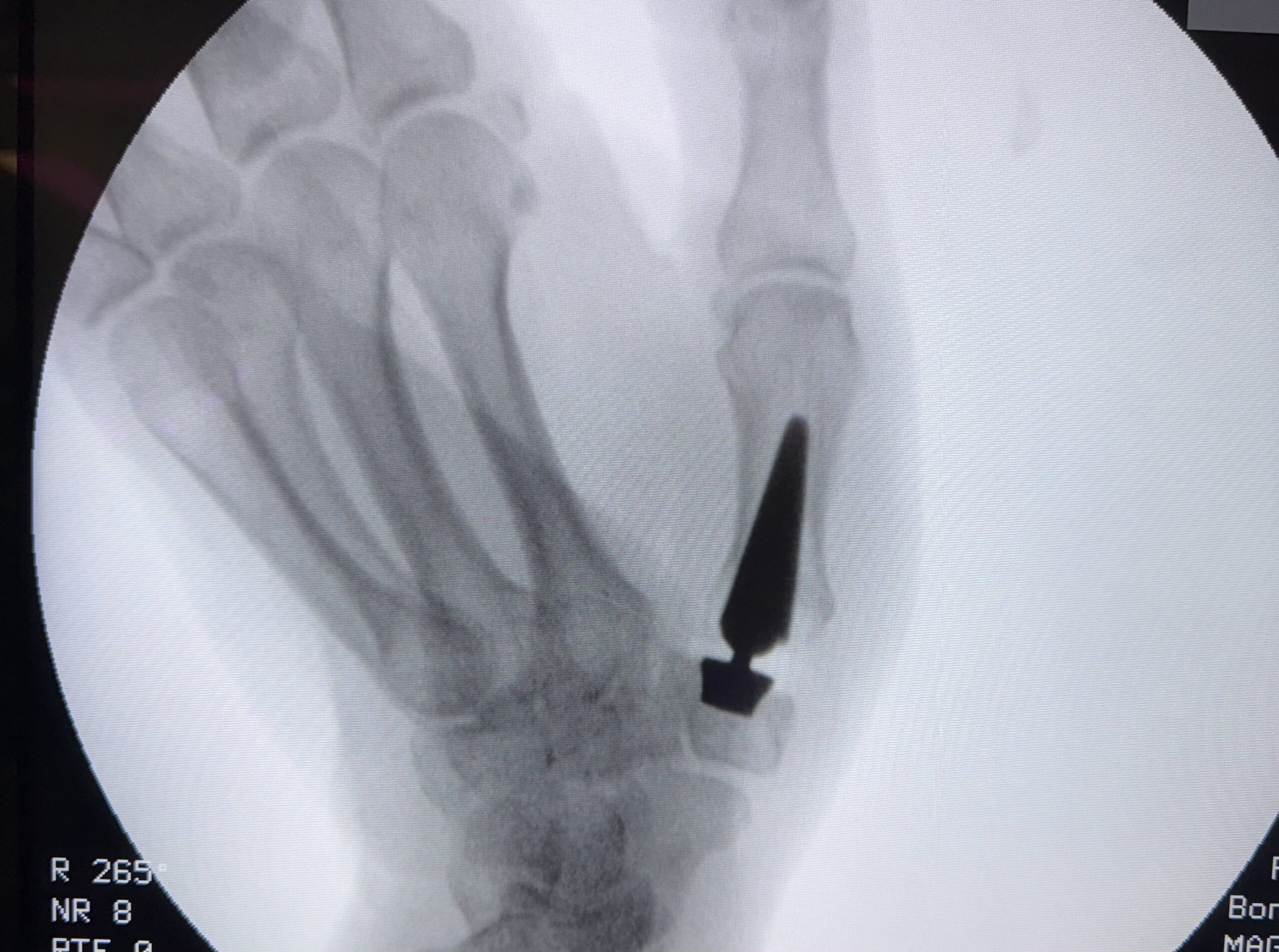
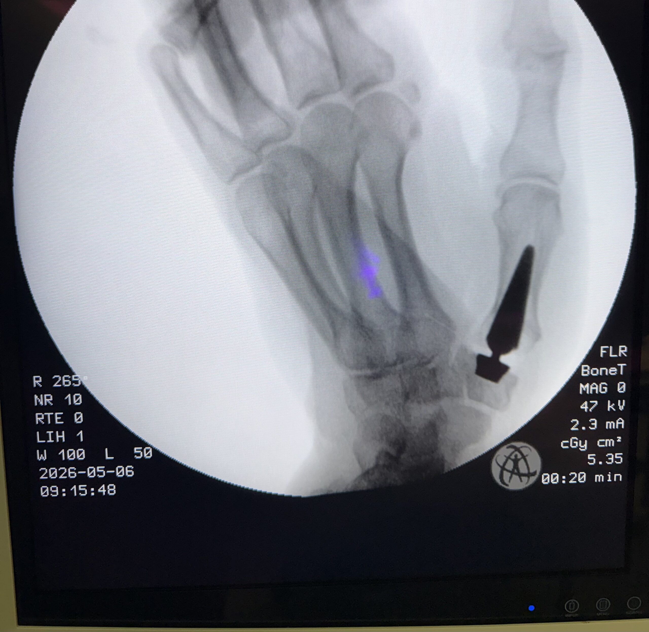
Thumb basal-joint arthritis is common, particularly in women over the age of 50. Traditional surgery often involves removing the arthritic trapezium bone. Although trapeziectomy can relieve pain, the first metacarpal may later settle into the space created by the excision, and grip strength may be reduced.
In selected patients, joint replacement may be considered. An artificial joint replaces the damaged carpometacarpal joint, with the aim of preserving thumb anatomy, stability and movement while allowing earlier mobilisation. The most appropriate treatment depends on the individual patient, the pattern of arthritis and functional requirements.
APTIS DRUJ replacement for distal radioulnar joint arthritis
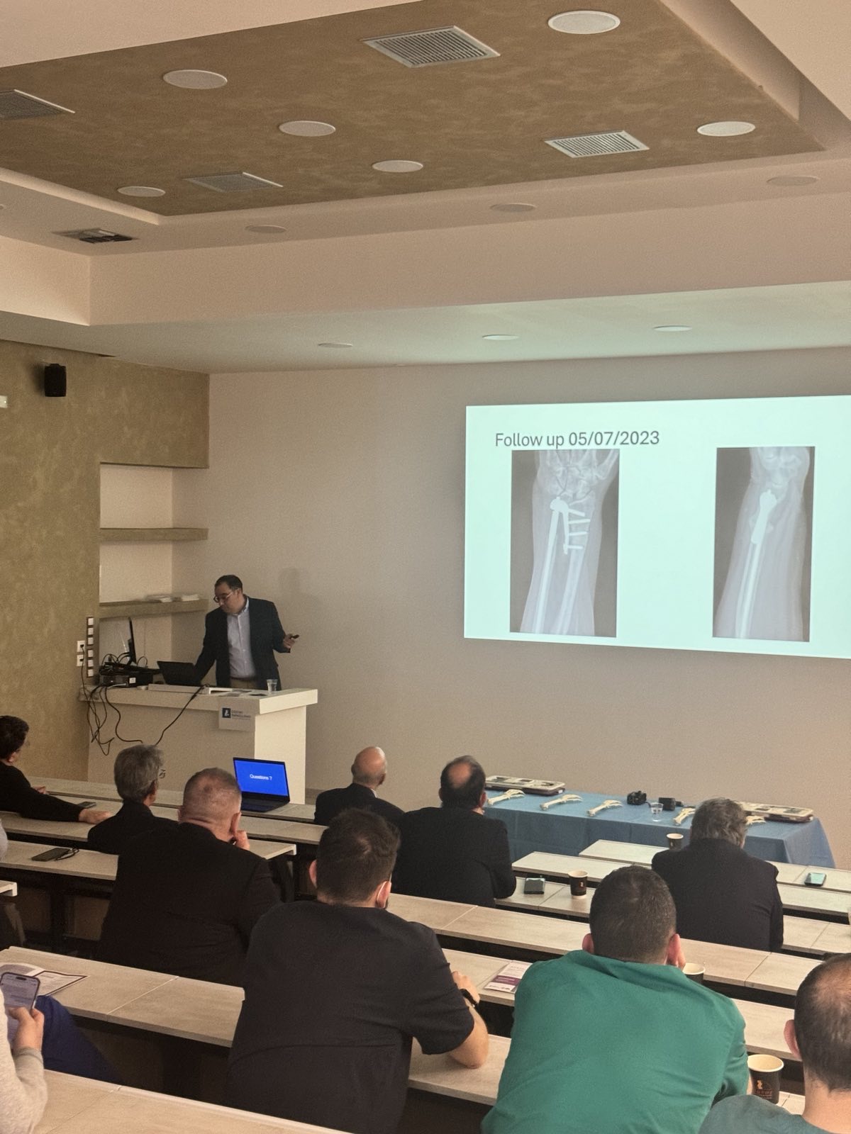
Dr Kritiotis introduced the APTIS DRUJ replacement in Cyprus in 2020. The distal radioulnar joint is the wrist joint that enables pronation and supination—the turning movement of the forearm.
Earlier salvage procedures for painful DRUJ arthritis included removal of the ulnar head (Darrach procedure) or fusion of the ulnar head to the distal radius with an osteotomy below the fusion (Sauvé-Kapandji procedure). In some patients, these procedures can result in forearm pain or weakness caused by an unstable ulnar stump.
The APTIS prosthesis is designed to stabilise the ulna relative to the radius while preserving forearm rotation. Developed by Dr Luis Scheker in Kentucky in the early 2000s, it is a specialist procedure performed by selected upper-limb surgeons worldwide.
Wrist arthroscopy
Dr Kritiotis has used wrist arthroscopy for diagnostic and therapeutic procedures since 2014. Arthroscopy enables the inside of the wrist joint to be examined through small incisions and can identify ligament injuries and other intra-articular conditions.
Procedures that may be performed arthroscopically include:
- Excision of persistent or recurrent wrist ganglia
- Repair or debridement of triangular fibrocartilage complex (TFCC) tears
- Selected ligament repairs
- Excision of osteophytes or early arthritic changes
- Excision and biopsy of benign intra-articular tumours
- Assessment and assistance during correction of complex or malunited distal-radius fractures
Correction of malunited fractures and deformities using 3D printing
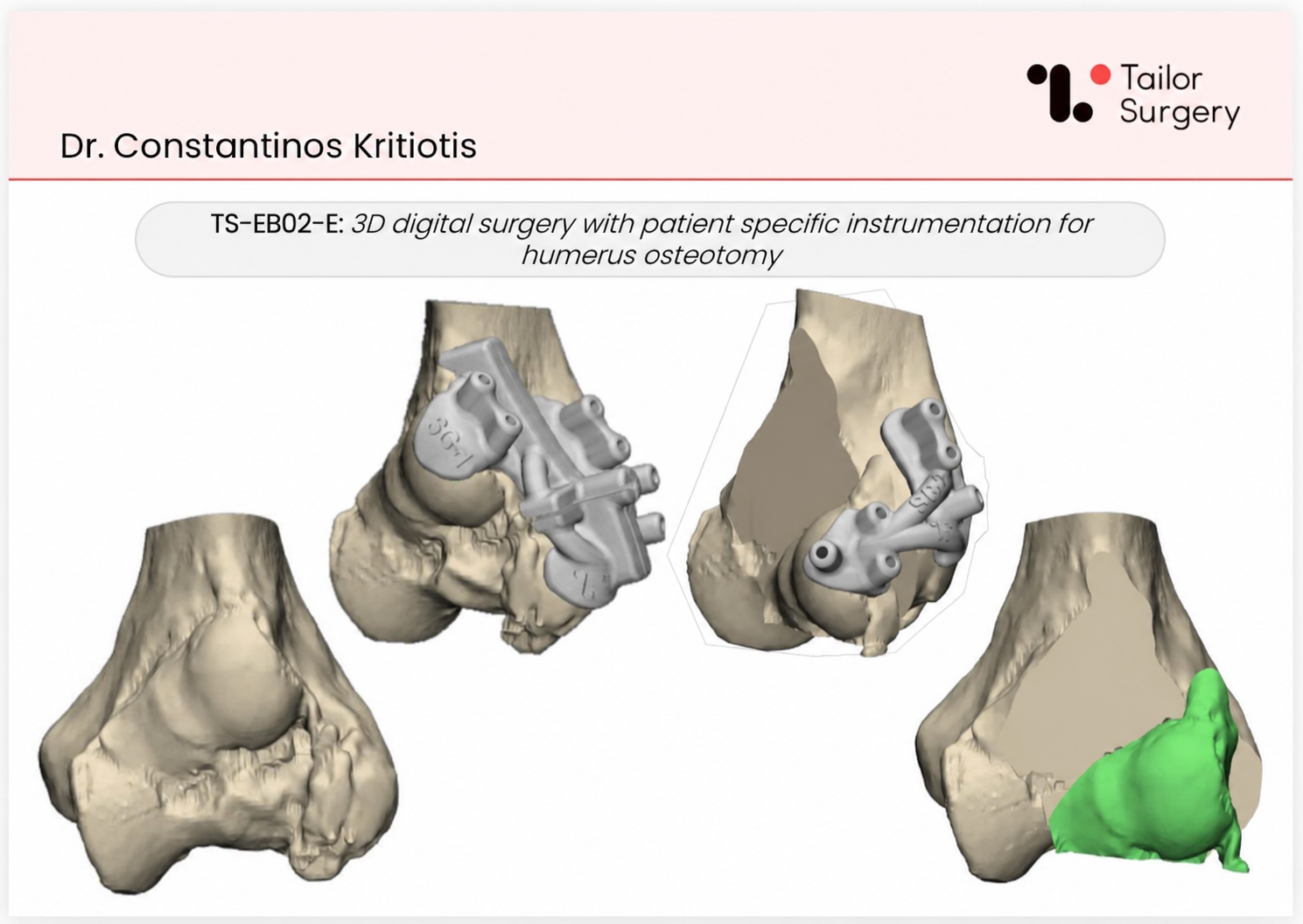
Dr Kritiotis has used patient-specific 3D-printed guides to correct malunited fractures of the distal humerus and wrist. CT scans of the affected and unaffected sides are used to plan the correction. Specialist software assists in planning the osteotomy, after which patient-specific cutting or positioning guides are printed and sterilised.
This process aims to reproduce the surgical plan as accurately as possible and restore the patient’s anatomy. Dr Kritiotis operates a small 3D-printing facility with filament and resin printers for surgical planning and guide production.
Acute hand trauma
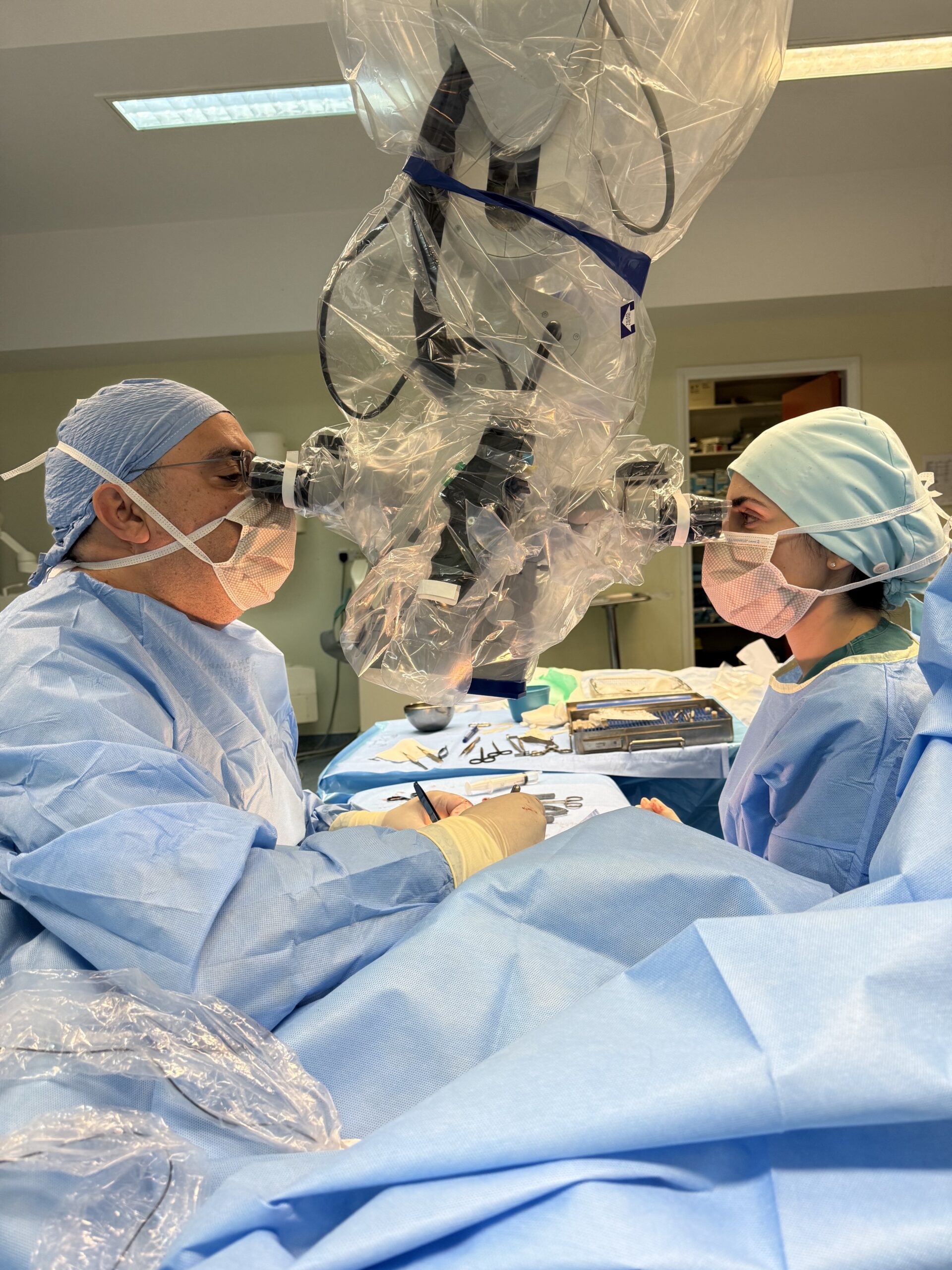
Dr Kritiotis provides a 24-hour service for acute hand-trauma cases, including complex lacerations involving tendons, nerves or blood vessels, as well as selected replantation cases. The operating theatre is equipped with a Zeiss operating microscope, an Orthoscan Mini C-arm, fracture-fixation systems and dedicated S&T microsurgical instruments.
Microsurgery
Tennis elbow (lateral epicondylitis)
Tennis elbow is an overuse condition affecting the tendons on the outer aspect of the elbow. Pain may radiate down the forearm and commonly worsens with gripping or lifting. Despite its name, it is more often caused by repetitive everyday activity than by sport.
Most people improve with rest, activity modification, physiotherapy, bracing and appropriate anti-inflammatory treatment. Surgery is rarely required and is reserved for persistent symptoms that have not responded to a suitable period of non-operative care.
Lacertus syndrome
Lacertus syndrome is compression of the median nerve beneath the lacertus fibrosus at the elbow, rather than at the wrist as occurs in carpal tunnel syndrome. Patients may experience reduced grip and pinch strength, hand fatigue and forearm pain, sometimes without numbness. It may therefore be mistaken for tennis elbow or a cervical-spine problem.
Diagnosis is primarily clinical because imaging and electrodiagnostic tests may be normal. Examination looks for characteristic muscle weakness and tenderness over the lacertus. Many patients improve with activity modification, taping and nerve-gliding exercises. When symptoms persist, release of the lacertus fibrosus can be performed through a small incision under local anaesthesia, allowing strength and symptoms to be assessed immediately.
Radial tunnel syndrome
Radial tunnel syndrome is compression of the posterior interosseous nerve as it passes through the proximal forearm, frequently near the arcade of Frohse. It can mimic tennis elbow, but the tenderness is usually several centimetres farther down the forearm and may worsen with resisted middle-finger or wrist extension and forearm rotation.
Symptoms are often predominantly painful, without numbness, although significant weakness of the finger and thumb extensors may occur in some cases. Diagnosis is mainly clinical, as imaging and electrodiagnostic studies may not demonstrate the compression. Initial treatment includes rest, splinting, activity modification and physiotherapy; persistent cases may benefit from surgical decompression.
Distal biceps tendon avulsion
A distal biceps tendon avulsion occurs when the tendon tears away from its attachment on the radial tuberosity. It most commonly affects men in their forties or fifties during a sudden eccentric load, such as attempting to catch a falling heavy object. Patients may feel a pop and sharp pain at the front of the elbow, followed by bruising and retraction of the muscle.
Complete tears reduce elbow-flexion strength and, more significantly, forearm supination strength. Diagnosis is usually clinical and may be supported by MRI when required. Because the tendon retracts and scars over time, surgical repair—when indicated—is generally best undertaken promptly, ideally within the first few weeks after injury.
Tumours and masses of the hand
Most lumps and bumps in the hand are benign. Common examples include ganglion cysts, giant-cell tumour of the tendon sheath and enchondroma. A giant-cell tumour of the tendon sheath is usually a slow-growing, firm mass adjacent to a tendon sheath or joint. An enchondroma is a benign cartilage tumour within a bone and may be discovered incidentally or following a fracture through the weakened bone.
Assessment combines clinical examination with appropriate imaging, such as ultrasound or MRI for soft-tissue masses and radiographs for bone lesions. Treatment depends on the diagnosis and symptoms and may include excision or curettage. Any lesion that grows rapidly, is painful or feels fixed to deeper structures should be assessed to exclude the uncommon possibility of malignancy.
Soft-tissue coverage of the hand
Soft-tissue defects can follow trauma or excision of a skin lesion. Because exposed tendon, bone, joint or nerve cannot usually be covered successfully with a simple skin graft, reconstructive flaps are often required to protect deeper structures and restore useful hand function.
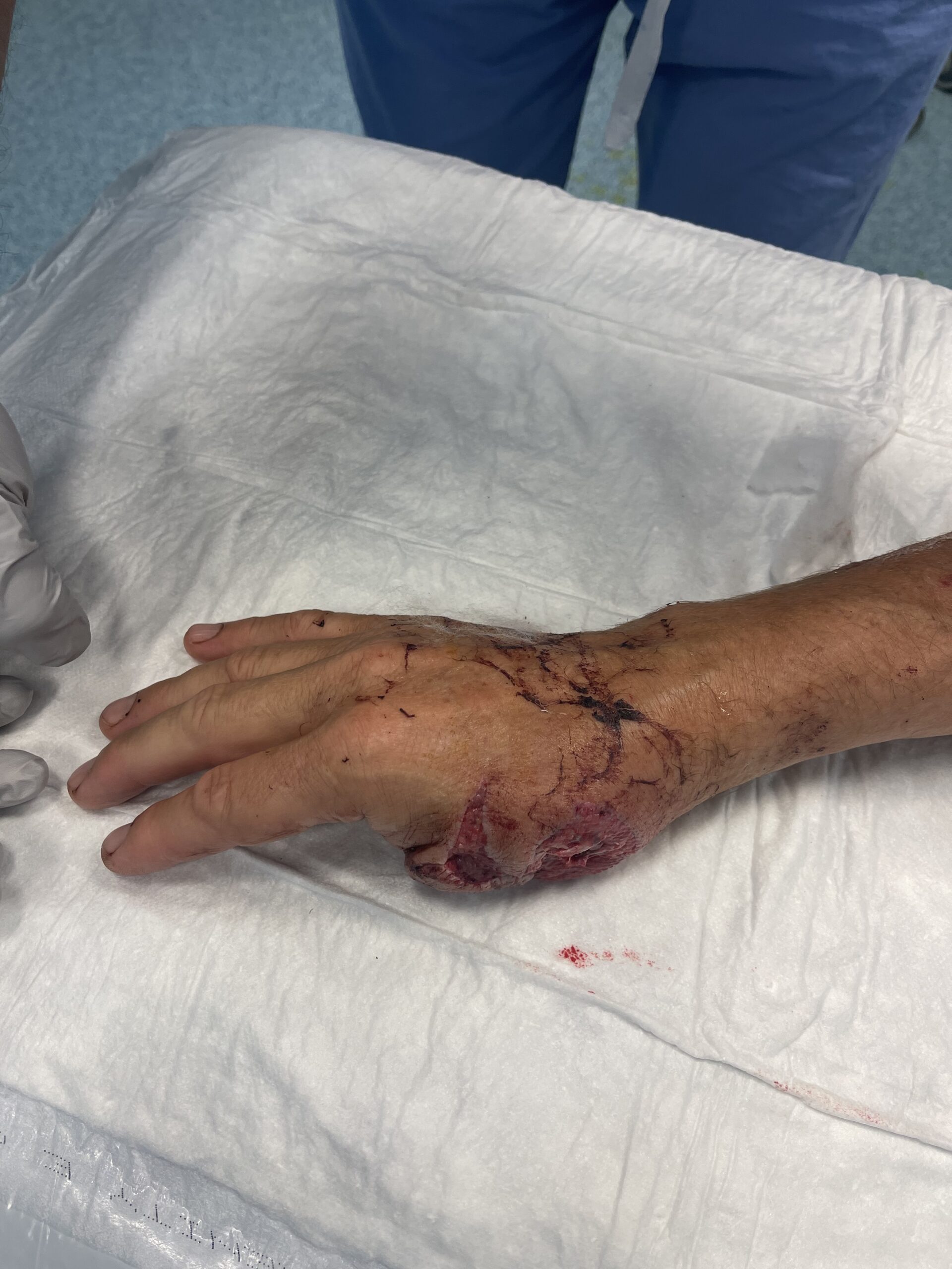
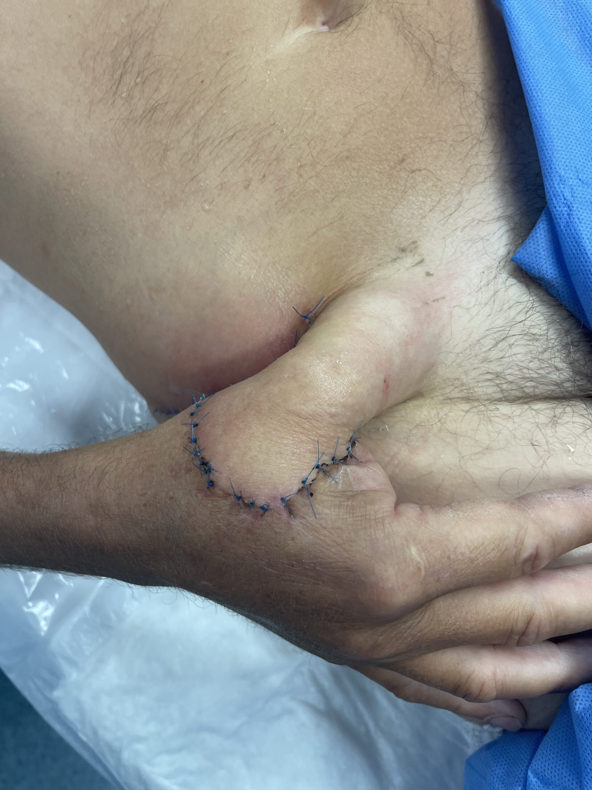
Small, well-vascularised defects—particularly at the fingertips—may be reconstructed with local flaps such as cross-finger, homodigital island, V-Y advancement, Moberg or first dorsal metacarpal artery flaps. Larger defects over the back of the hand or wrist may require regional pedicled flaps, including posterior interosseous artery or reverse radial forearm flaps.
When a defect is extensive, contaminated or unsuitable for local and regional options, free-tissue transfer can provide durable, well-vascularised coverage. The choice of reconstruction balances the size and location of the defect, the structures requiring protection, donor-site effects and the patient’s functional needs.